Vision & Imaging Blog
Microscopy in Life Sciences Used to Track Bee Pollination Processes
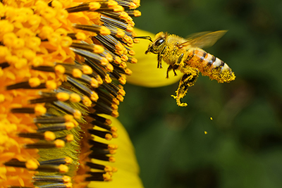 Innovative use of quantum dots, nanocrystals of semiconductor materials that are so small that they behave like artificial atoms, and the fluorescence microscope finally may be the solution scientists have been looking for to track pollination from start to finish. Researches still don’t know where most microscopic pollen grains land once they leave flowers. Which pollinators transfer the grains? And where do they come from? This new, cost-effective method can help track whether there was a successful transfer or if the pollen grain was lost along the way.
How Fluorescence Stereomicroscopes Work
Fluorescence microscopes use fluorescence and phosphorescence instead of (or in addition to) reflection and absorption to study properties of a substance. The stereo fluorescence illuminates a sample with a light with the wavelength that excites fluorescence in the sample.
The main task of a fluorescence microscope is to let excitation light radiate the specimen. It then sorts out the weaker emitted light from the image. The light is imaged through the microscope objective and all the light except the desired wavelength is filtered out.
ROI Calculator

Discover the potential cost savings of robotic automation over a 20-year system life
This calculator compares your current manual labor costs against the total cost of owning and operating a robotic system over its 20-year lifespan.
Researchers Find a New Way to Track Pollen Grains
Inspired by the use of quantum dots used to track cancer cells in rats, researchers came up with an advanced technique to keep tabs on pollen grains. When quantum dots are exposed to UV light, they fluoresce extremely bright light in the visible and IR wavelengths. They can be used as alternatives to organic dyes. Quantum dots with lipophilic ligands stick to the fatty outer layer of pollen grains, called pollenkitt. The glowing colors could then be used to label the pollen grains and see where they end up.
Researches used nontoxic quantum dots with ligands as pollen-grain labels. Using a micropipette, they dispensed quantum dots dissolve in hexane directly onto dehisced anthers. The hexane solvent evaporates and leaves the quantum dots attached to pollen grains. They first tested to make sure that honeybees transferred the labeled grains no differently than unlabeled pollen grains.
The next step was to find a way to view the fluorescing pollen grains under a field dissection microscope. Researchers used a fluorescence box that fits under a fluorescence microscope. The box can be 3D-printed at a relatively low cost. This innovative process used existing microscopy technology to help develop a low-cost, effective solution to the pollen tracking problem.
Share This On X:
Recent Posts
- How 3D Vision Systems Are Transforming Food Manufacturing
- Autonomous Driving Technology: Dueling Approaches Fight for Control of the Wheel
- These Farms Are Harnessing Machine Vision for Smarter Agriculture
- Revealing the Hidden Effects of Climate Change with Advanced Imaging
- How Vision Systems Are Transforming Predictive Neurodiagnostics
- Innovative Machine Vision Lenses and Trends
- View All Vision & Imaging Blogs