Vision & Imaging Blog
High-Speed 3D Microscope Creates Images of Living Creatures in Real-Time
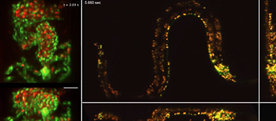 In early 2019, a high-speed, 3D microscope technology called SCAPE first offered engineers and neuroscientists the chance to see individual nerve cells moving, stretching, and switching on and off. The data collected about how neurons work together will help researchers understand the secrets behind how the brain responds to stimuli and how it organizes information.
Collaborating with scientists around the world, researchers have improved the SCAPE technology to produce faster, 3D images that should impact fields like genetics, cardiology, and neuroscience. The team claims that the improved version can capture images up to 30 times faster than they originally demonstrated earlier this year.
Why Scientists Built the SCAPE 3D Microscope
Living things are dynamic. From one moment to the next, an animal’s cells make significant changes. Faster 3D microscope imaging lets scientists observe a being’s biological processes as a whole and how they interact with one another. SCAPE, which stands for Swept Confocally Aligned Planar Excitation, was its development team’s solution to the limits of traditional microscopy.
Traditional 3D microscopes used to image living samples can only scan a small spot of laser light around the sample. The approach is slow and only allows for a short time to see the spot. SCAPE uses a sheet of angled light to illuminate an entire plane within a sample. It then sweeps the light across the sample to form a 3D image. SCAPE’s design makes it much faster than traditional 3D microscopes. Since SCAPE uses a single lens, it’s also simple and compact.
ROI Calculator

Discover the potential cost savings of robotic automation over a 20-year system life
This calculator compares your current manual labor costs against the total cost of owning and operating a robotic system over its 20-year lifespan.
SCAPE 2.0 Improves the Already Amazing 3D Microscope
Advances in fluorescent labeling have increased overall interest in the SCAPE 3D microscope technology. These advances let scientists make specific cells in an animal glow in different colors. The cells can even flash on and off when signaling each other. SCAPE 2.0 can capture cellular events in vivid detail, including the movements and responses that occur as a result.
Since the 3D microscope’s initial release, scientists have used near-transparent animals like worms, zebrafish embryos, and fruit flies to study natural behaviors and have even modified them to emulate the behavior of human diseases. One cardiologist even used SCAPE within a zebrafish model to observe how genetic mutations can cause the heart to be malformed. The doctor hopes to find a link between such mutations and children living with congenital heart disease. The 3D microscope only appears to be limited by what scientists can dream of studying.
Recent Posts
- How 3D Vision Systems Are Transforming Food Manufacturing
- Autonomous Driving Technology: Dueling Approaches Fight for Control of the Wheel
- These Farms Are Harnessing Machine Vision for Smarter Agriculture
- Revealing the Hidden Effects of Climate Change with Advanced Imaging
- Innovative Machine Vision Lenses and Trends
- An Early Look at GigE Vision 3.0
- View All Vision & Imaging Blogs