Robotics Blog
Researchers Leverage Two Photon Microscopy and Mesoscopic Imaging to Capture Real-Time Brain Activity
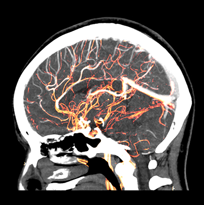 Yale scientists have combined two technologies, two-photon microscopy and mesoscopic imaging, to capture real-time brain activity for the first time. Until now, scientists had to speculate about the complex interactions across the brain. This new imaging technique offers a groundbreaking view of the brain at work in real-time, giving researchers an overall view with detailed data of what is actually happening.
Researchers hope to be able to use the data to track learning and cognitive processing, first starting with mice. More testing is needed to determine if the procedure is safe enough to be used on human subjects. Because basic brain architecture and function are similar across all mammals, observation of the mouse’s brain will give scientists in-depth information on how the human brain operates as well.
Combining Two Photon Microscopy and Mesoscopic Imaging
The multi-lab collaboration, funded by the National Institutes of Health BRAIN Initiative, created the method that uses a microscope with an orthogonal axis design. The mesoscopic objective is oriented above the subject’s brain and the two-photon objective is oriented horizontally, with imaging performed through a micro prism.
ROI Calculator

Discover the potential cost savings of robotic automation over a 20-year system life
This calculator compares your current manual labor costs against the total cost of owning and operating a robotic system over its 20-year lifespan.
Before developing this technique, scientists typically only captured images that focused on single molecules, cells, or circuits. This new method captures the behavior of both individual neurons and the entire brain. With this development, scientists now can see what individual neurons are doing in relation to the rest of the brain.
New Imaging Technique Improves Upon Existing Methods
Scientists currently use Functional Magnetic Resonance Imaging (fMRI) scans to gather data on how the brain works. But fMRI only monitors where blood is flowing within the brain and is more of an indirect readout of brain activity. The two photon microscopy method is much more detailed and can analyze the brain’s activity much faster. Currently, fMRI monitors brain activity in a matter of seconds, but this new technique can capture brain activity in milliseconds.
Researchers hope to use this information to understand better how different parts of the brain affect emotions and critical thinking. Ideally, this new imaging technology also could potentially be used to study how diseases like Alzheimer’s develop.
Scientists will be able to use the data to see how brain circuits change when animals learn. This may offer insight into neurodevelopmental diseases like autism. With more understanding about how the brain works, more effective medicines and therapies can be developed to fix its problems.
Recent Posts
- How 3D Vision Systems Are Transforming Food Manufacturing
- Autonomous Driving Technology: Dueling Approaches Fight for Control of the Wheel
- These Farms Are Harnessing Machine Vision for Smarter Agriculture
- Revealing the Hidden Effects of Climate Change with Advanced Imaging
- How Vision Systems Are Transforming Predictive Neurodiagnostics
- Innovative Machine Vision Lenses and Trends
- View All Robotics Blogs