Vision & Imaging Blog
Machine Vision in Life Sciences: Using an Electron Microscope to Create High-Res 3D Images of Cells
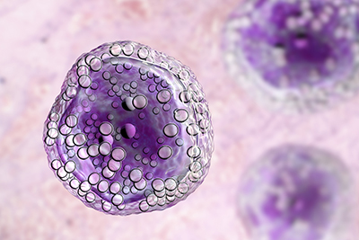 3D machine vision is helping researchers find new drugs to combat diseases and see how those drugs work at a cellular level. Many new drugs fail when they reach clinical trials because their effects are not fully understood. But 3D images of cells created with the use of an electron microscope can help scientists further understand disease and revolutionize how new medicines are developed.
Improving Cryogenic Electron Tomography
Cryogenic Electron Tomography (cryo-ET) builds up a 3D image from multiple 2D images of cell samples frozen at temperatures below -180°C (Celsius). Presently, cryo-ET can only handle small samples, such as parts of cells. But researchers are looking to speed up the technique to process much larger samples.
New techniques will need to be developed to prepare and handle samples that are less than 1/100ththe thickness of a human hair. The new electron microscopy techniques will also have to speed up the imaging process and collect the huge amount of data created. In addition to these challenges, new software and machine learning technology will need to process the data to create and interpret 3D images.
ROI Calculator

Discover the potential cost savings of robotic automation over a 20-year system life
This calculator compares your current manual labor costs against the total cost of owning and operating a robotic system over its 20-year lifespan.
Improvement in patient biopsies, increased workflow automation, and the standardization of post-processing data are the focused areas of development. The long-term goal is to bring the technology closer to the clinic so it can be used in research hospitals and laboratories.
Electron microscopy can help researchers study the role of genes and proteins as part of a whole cell or a group of cells. Experts can see the molecular processes that are set in motion by a genetic mutation.
Machine Vision Used to Create 3D Models
The collection of data by use of 3D machine vision and electron microscopy is helping experts to create 3D models to test new medicines. One major pitfall when translating drugs from the lab to a viable treatment is determining the differences from the 2D petri dish to the 3D environment of the human body.
Researchers have used a new 3D bioprinter to mimic the composition of physiological structures and let cells behave similarly to when they are in the human body. 3D bioprinting lets researchers place cancer cells, normal cells, and blood vessel structures in the lab dish.
Fluorescence imaging then lets them observe the way the cancer cells migrate. Because the 3D models are bioprinted, it is a reproducible process that can be used to quickly run thousands of tests. Researchers hope this model will help them understand how cancer spreads and how to create better treatments.
Recent Posts
- How 3D Vision Systems Are Transforming Food Manufacturing
- Autonomous Driving Technology: Dueling Approaches Fight for Control of the Wheel
- These Farms Are Harnessing Machine Vision for Smarter Agriculture
- Revealing the Hidden Effects of Climate Change with Advanced Imaging
- Innovative Machine Vision Lenses and Trends
- An Early Look at GigE Vision 3.0
- View All Vision & Imaging Blogs