Blog
How Vision Systems Are Transforming Predictive Neurodiagnostics
 Neurodegenerative diseases such as Alzheimer’s (AD) and Parkinson’s (PD) represent some of the most complex and pressing challenges in global healthcare. These disorders are marked by the progressive and irreversible loss of neurons, resulting in cognitive, motor, and behavioral impairments that worsen over time. What makes them particularly insidious is their prolonged asymptomatic phase: pathological changes can unfold silently over the course of decades before clinical symptoms surface. By the time memory lapses, tremors, or speech difficulties become noticeable, the underlying neurological damage is often extensive and irreversible.
This delayed onset poses a fundamental barrier to effective intervention. With no existing cures and a rapidly aging global population, the medical community is shifting its focus from reactive care to predictive diagnostics. Emerging tools at the intersection of advanced imaging and machine intelligence are now enabling a deeper, earlier understanding of how neurodegeneration begins. Vision systems powered by AI are revealing subtle biomarker patterns and structural changes in the brain well before cognitive decline is measurable, creating new possibilities for timely intervention and long-term disease management.
Imaging the Invisible: The Rise of Predictive Diagnostics
Conventional diagnostics for Alzheimer’s and Parkinson’s rely heavily on subjective cognitive assessments and observable symptoms. However, these methods are inherently reactive. Modern biomedical imaging, empowered by artificial intelligence (AI), is redefining this paradigm through predictive diagnostics, enabling clinicians to identify early neural deviations that precede clinical manifestation by years or even decades.
At the heart of this transformation are imaging modalities such as positron emission tomography (PET), functional magnetic resonance imaging (fMRI), retinal fundus imaging, and multi-photon optical microscopy. When coupled with AI-powered vision systems, these tools enable deep phenotyping of neurological tissue, uncovering subtle patterns of degeneration invisible to the human eye.
For example, PET imaging has proven critical in tracking amyloid-beta accumulation and tau protein tangles, which are hallmarks of Alzheimer’s disease, long before memory loss or behavioral changes occur. Similarly, functional magnetic resonance imaging (fMRI) captures real-time disruptions in neural connectivity, a growing biomarker in Parkinson’s disease research. These insights are only possible through sophisticated imaging algorithms capable of processing high-resolution, high-volume neural data with extreme precision and speed.
How Do AI Powered Digital Biomarkers Detect Parkinson’s Early?
Traditional biomarkers, such as cerebrospinal fluid analysis or structural imaging, face practical limitations. They are invasive, expensive, and often fail to detect the earliest signs of disease. In contrast, digital biomarkers represent a non-invasive, scalable alternative. Powered by machine vision and AI, they collect continuous, high-frequency data from various sources: retinal scans, facial microexpressions, eye movements, and gait patterns to assess neurocognitive function.
Recent studies have demonstrated how convolutional neural networks (CNNs) trained on eye-tracking data can distinguish between PD patients and healthy controls with ROC-AUC scores as high as 0.88. Similarly, facial expression analysis tools have achieved diagnostic accuracies approaching 90 percent, identifying emotional flattening commonly associated with neurodegeneration. These models can operate passively, remotely, and in real time, opening the door to widespread population screening without the need for traditional hospital infrastructure.
Retinal imaging, in particular, has emerged as a powerful window into neurodegeneration. The retina, an extension of the central nervous system, exhibits microvascular changes and structural degradation reflective of cerebral pathology. In a groundbreaking application, AI-based analysis of fundus images from the UK Biobank predicted the incidence of Parkinson’s disease up to seven years before diagnosis, with sensitivities reaching over 90 percent in key time windows. Deep neural networks were able to identify critical retinal features including optic disc cupping, vascular displacement, and foveal contour shifts, all of which are impossible to quantify manually.
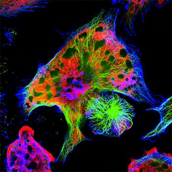
Optical Microscopy and the Power of Resolution
While digital biomarkers provide scale and accessibility, advanced optical imaging delivers the resolution and spatial fidelity required to interrogate the brain’s structural and functional architecture at the most granular levels. Techniques such as two-photon and three-photon microscopy, fluorescence imaging, and light sheet microscopy offer researchers the ability to visualize dynamic cellular processes within intact tissue environments. These modalities are particularly critical for mapping synaptic degradation, neuroinflammation, and alterations in neurovascular coupling, which are all core mechanisms driving the onset and progression of Alzheimer’s and Parkinson’s diseases.
At the forefront of this imaging frontier is the mesoSPIM (mesoscale selective plane-illumination microscope), a platform engineered to overcome the limitations of traditional sectioning and confocal microscopy. By employing light-sheet illumination for optical sectioning, mesoSPIM generates ultra-high-resolution, volumetric reconstructions of entire brain regions without inducing mechanical damage. This approach preserves tissue integrity while dramatically accelerating acquisition speed. As a result, researchers can conduct longitudinal studies of neurodegeneration with unprecedented anatomical clarity, enabling the precise characterization of disease progression, network reorganization, and therapeutic impact over time.
ROI Calculator

Discover the potential cost savings of robotic automation over a 20-year system life
This calculator compares your current manual labor costs against the total cost of owning and operating a robotic system over its 20-year lifespan.
Vision Systems and AI: Engineering Challenges in Medical Practice
Despite these technological leaps, integrating AI-driven vision systems into clinical workflows remains a complex undertaking. Translational gaps persist, particularly in standardization, regulatory validation, and interoperability with existing health systems. Models trained on narrow datasets often fail in real-world settings due to population variance, comorbidities, or imaging inconsistencies.
To overcome these challenges, future solutions must embrace robustness at three critical levels:
- Disease-specific modeling: AD and PD have heterogeneous subtypes and overlapping pathologies. AI models must adapt to these nuances while preserving diagnostic specificity.
- Task-specific optimization: Feature extraction pipelines need to be tailored for classification, progression modeling, or therapeutic monitoring, each with distinct data demands.
- Technology-specific resilience: From cloud orchestration to edge computing in diagnostic equipment, system architectures must support real-time inference, data security, and explainability for regulatory compliance.
The case of Google’s Automated Retinal Disease Assessment (ARDA) system illustrates this complexity. Though successful in research settings, the tool struggled with deployment in clinical environments lacking infrastructure and cultural alignment. For neurodegenerative disease imaging to avoid similar pitfalls, cross-disciplinary collaboration will be essential, linking radiologists, data scientists, hardware engineers, and ethicists in a unified pipeline.
What Does Machine Vision Mean for Next-Gen Neurocare?
The convergence of machine vision and predictive diagnostics is unfolding now. Medical imaging is one of the most active domains for AI integration, with vision-based diagnostics leading clinical AI deployments across hospitals and research centers worldwide. This reflects a broader shift toward precision, speed, and scalability in neurological care. As PET, fMRI, and optical microscopy evolve alongside digital biomarkers and AI models, the vision systems powering neurodegenerative diagnostics are becoming more accurate, less invasive, and more accessible.
This shift could redefine how society approaches brain health. Instead of waiting for irreversible cognitive decline, clinicians will have the tools to intervene in the silent years of neurodegeneration, extending quality of life, reducing economic burden, and enabling more targeted therapeutic strategies.
For professionals at the intersection of vision technology, machine learning, and life sciences, this is a moment of unprecedented impact. The ability to image, model, and understand the brain with precision was once confined to theoretical ambition. Today, it is becoming a clinical imperative.
Advancing the Future of Neurodiagnostics Through Vision and AI
Imaging and vision technologies are actively redefining the boundaries of early diagnostics in neuroscience and biomedical science. As these systems scale, safeguarding patient privacy and ensuring ethical AI deployment will be critical components of their design. The Association for Advancing Automation (A3) remains deeply embedded in the research and innovation driving this transformation. From enabling next-generation optical microscopy platforms to supporting the integration of AI into clinical imaging workflows, A3 serves as a vital nexus between academic research, industry pioneers, and automation professionals. Our members and partners are at the forefront of developing high-resolution, high-throughput vision systems that are actively reshaping the future of neurodegenerative disease detection and care.
Whether you're exploring digital phenotyping for Alzheimer’s, deep learning for Parkinson’s diagnostics, or the hardware-software interface in medical imaging, A3 offers direct access to the thought leaders and technologies driving these breakthroughs.
Explore what’s next in vision systems, biomedical imaging, and intelligent diagnostics. Subscribe to A3’s monthly newsletters for expert insights, sector-specific use cases, and exclusive coverage of the technologies shaping the future of neuroscience, healthcare, and machine intelligence. You’ll be well-positioned for what comes next.
Glossary
PET Imaging: Positron emission tomography, a nuclear medicine technique that uses a small amount of radioactive tracer to measure brain metabolism, blood flow, and chemical activity. For Alzheimer’s and Parkinson’s, it can identify early disease markers like amyloid plaques, tau tangles, or reduced dopamine activity before symptoms appear, enabling earlier and more accurate diagnosis.
Retinal Imaging: The use of high-resolution imaging technologies, such as optical coherence tomography or fundus photography, to capture detailed images of the retina. For Alzheimer’s and Parkinson’s, it can reveal early signs of neurodegeneration through changes in retinal structure or blood vessels, offering a noninvasive way to aid early detection and disease monitoring.
Digital Biomarkers: Objective, quantifiable data collected through digital devices such as wearables, smartphones, or sensors. For Alzheimer’s and Parkinson’s, they can track subtle changes in movement, speech, cognition, or daily activity patterns over time, helping detect disease earlier and monitor progression more precisely.
Share This On X:
AI + imaging = a new frontier in brain diagnostics. See how PET, fMRI, and retinal scans are detecting Alzheimer’s and Parkinson’s before symptoms emerge. #AIinMedicine #Neurodiagnostics #BrainHealth
Digital biomarkers and deep learning are unlocking pre-symptomatic detection of neurodegenerative disease. Explore the tools leading the charge. #Neuroscience #DigitalHealth #PredictiveAI
Recent Posts